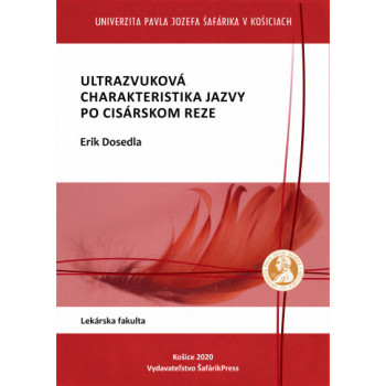
Ultrazvuková charakteristika jazvy po cisárskom reze

E-book

Erik Dosedla

A uterine scar is currently a significant medical problem. Uterine scars most commonly occur after a Cesarean section, which is one of the most frequent laparotomic surgeries (Yazicioglu et al., 2006). The frequency of Cesarean sections increased between 1990 and 2014 in the USA from 21.2% to 32.2% (Hamilton et al., 2015), in Great Britain from 12% to 26.2% (NHS Maternity Statistics - England, 2013-14: Summary Report, 2015), and in the Slovak Republic from 8.7% to 31% (Národné centrum zdravotníckych informácii, 2016).

After a Cesarean section (CS), there is a risk of abnormal healing of the uterotomic wound, which can result in the development of certain pathological conditions in consecutive pregnancies (uterine rupture, ectopic pregnancy in the Cesarean scar, placental disorders) (Seow et al., 2004; Oyelese and Smulian, 2006; Fitzpatrick et al., 2012). In addition, a defect in the Cesarean scar is associated with chronic dysmenorrhea, chronic pelvic pain, infertility, and postmenstrual spotting (Wang et al., 2009; Bij de Vaate et al., 2011).

Ultrasound is a valuable examination method in the diagnostic and therapeutic management of these pathological conditions.

Download e-book for free (pdf)

Quantity

978-80-8152-908-5

Data sheet

Method of publication:
E-book (pdf)
Author:
Erik Dosedla
Document type:
Monograph
Number of pages:
83
Available from:
20.10.2020
Year of publication:
2020
Edition:
1st edition
Publication language:
Slovak
Faculty:
Faculty of Medicine
License:
CC BY NC ND (Uveďte autora - Nepoužívajte komerčne - Nespracovávajte)
- Free for download

16 other products in the same category:

Multidisciplinárny pohľad na pacienta s...

€21.00
Availability: 19 In Stock

Martina Zavacká a kolektív

Kniha „DIABETICKÁ NOHA Z POHĽADU CIEVNEHO CHIRURGA“ obsahuje 14 kapitol. Pojednáva o syndróme diabetickej nohy z multidisciplinárneho pohľadu viacerých odborníkov: angiochirurga, diabetológa, chirurga, podiatra, röntgenológa, neurológa a algeziológa. Úvodné kapitoly majú charakter propedeutiky, čo má taktiež význam aj pre pregraduálnu výchovu. Nasledujúce kapitoly sa zaoberajú vznikom syndrómu, výskytom, jeho diagnostikou, terapeutickými postupmi, pridruženými postihnutiami neurologického charakteru, starostlivosťou podiatra a algeziológa pri pretrvávaní bolestí. Ďalšie časti sú vyhradené chirurgickej a endovaskulárnej liečbe tepien cestou cievneho chirurga a intervenčných angiológov. Nezabúdajúc na problematiku amputácií a správnu rehabilitáciu pacientov s diabetom.  V knihe nájdu poučenie cievni chirurgovia, angiológovia, všeobecní chirurgovia, diabetológovia, neurológia, röntgenológovia, podiatri  a všeobecní lekári.

Kniha je zdrojom najnovších teoretických i praktických informácií pre postgraduálnu výchovu vyššie spomenutých odborníkov.

</

Pathology Part 1 Practical lesons for students...

€15.00
Availability: 100 In Stock

Alžbeta Blichárová a kolektív

“Pathology Part 1 (winter semester) - Practical lessons for students of general medicine” represents the first half of the university textbook designed for the pregradual study of pathology. The textbook complements the lectures of pathology and serves as a source of information for students of general medicine, that are preparing for the practical as well as the final oral examination.

In this part of the “Pathology” series, seventy selected histopathological slides are presented. Each selected slide is provided with general information in relation to etiology and pathogenesis of the disease, as well as with corresponding morphological features, including the gross appearance and the most important histopathological characteristics. The textbook summarizes the basic concepts of general pathology that are necessary to master the broad field of systemic pathology in the second semester.

Keywords: general pathology – histopathological slides – general medicine – gross appearance – microscopic image


Examination methods and injection technique in...

E-book

E-book

Ahmad GharaibehMarek Lacko

Authors describe the clinical and paraclinical examination methods necessary for the diagnosis and treatment of orthopedic diseases in both children and adults.

The examination methods are categorized based on individual anatomical locations. A valuable chapter discusses joint aspirations, detailing their indications, techniques for performing them, and potential complications. The chapter showcasing patient documentation for students serves as a significant aid for practical exams before the state final examination in surgery.

This work appropriately complements the available educational literature required for international undergraduate students in general medicine, as well as for physicians in specialized training in orthopedics and related fields.

Download e-book for free (pdf)

Základy histológie II. Učebnica a atlas...

E-book

E-book

Iveta Domoráková - Štefan Tóth - Zuzana Fagová - Eva Mechírová - Alexandra Kunová - Kristína Čurgali - Viera Eliášová - Monika Holodová - Marianna Danková

In our new electronic textbook, FUNDAMENTALS OF HISTOLOGY II – Textbook and Atlas of Microscopic Anatomy of Organs, we present students and the professional public with essential information about the microscopic structure of organs, accompanied by extensive visual documentation of organ structures and clear legends for the microphotographs.

All histological specimens come from the archive of the Institute of Histology and Embryology, are used in practical exercises, and are also included in teaching presentations. The electronic textbook is intended for undergraduate and postgraduate students of general medicine and dentistry at medical faculties, as well as for students of veterinary medicine and pharmacy, and biology students at faculties of natural sciences.

The textbook is designed so that students can find clear explanations of most medical terms and also reinforce their knowledge of professional terminology in Latin. An advantage of the electronic textbook is the ability to enlarge microscopic photographs, allowing detailed observation of structures without any qualitative loss of the viewed image. The microphotographs used in the atlas were taken with the following equipment: Zeiss Promicra and Olympus BX50 with a Canon EOS 2000D digital camera and QP Industrial 3.2 software.

Download e-book for free (pdf)

This website uses cookies to ensure you get the best experience on our website