There are 5 products.
Histology (Practical lessons)
€9.26
Availability: 50 In Stock
Eva Mechírová - Iveta Domoráková
The content of this "Practical lessons" represents a record of the appearance, under the light microscope of the tissues, organs and systems of the human body. In addition, it provides visual guidelines during the practical phase of a histology course. It is hoped that this book will be especially useful to students to understand fully the microscopic structure of human tissues and organs.
Authors
Vybrané kapitoly z histológie pre odbor zubného...
E-book
E-book
Iveta Domoráková et al.
The e-learning textbook is available at: https://portal.lf.upjs.sk. The textbook contains 11 chapters focusing on the microscopic anatomy of the head and neck region, with an emphasis on the oral and nasal cavities, skull bones and their development, and the detailed microscopic structure of teeth and their development. One chapter is dedicated to dentition from the perspective of the clinical significance of understanding tooth structure and development. The textbook includes chapters on the microscopic anatomy of organs that are functionally important for the head region, such as the vascular, lymphatic, endocrine, and nervous systems. The textbook is linked to an atlas, has a total of 490 pages, and features 300 original microphotographs using common histological stains, histochemical, immunohistochemical, and impregnation methods.
The textbook was created with the support of the KEGA project 019UPJŠ-4/2016.
Základy histológie I. Učebnica a mikroskopický...
E-book
E-book
Iveta Domoráková - Štefan Tóth - Zuzana Fagová - Kristína Čurgali - Viera Eliášová - Alexandra Kunová - Monika Holodová
The peer-reviewed textbook and microscopic atlas is aimed at understanding the basic microscopic structure of cells and tissues in a light microscope. Texts in the textbook are complemented by a rich set of histologic micrographs with basic and specific staining techniques.
All histologic micrographs were prepared by the author's team at the Department of Histology and Embryology, Faculty of Medicine, UPJŠ.
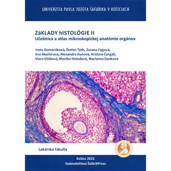
Základy histológie II. Učebnica a atlas...
E-book
E-book
Iveta Domoráková - Štefan Tóth - Zuzana Fagová - Eva Mechírová - Alexandra Kunová - Kristína Čurgali - Viera Eliášová - Monika Holodová - Marianna Danková
In our new electronic textbook, FUNDAMENTALS OF HISTOLOGY II – Textbook and Atlas of Microscopic Anatomy of Organs, we present students and the professional public with essential information about the microscopic structure of organs, accompanied by extensive visual documentation of organ structures and clear legends for the microphotographs.
All histological specimens come from the archive of the Institute of Histology and Embryology, are used in practical exercises, and are also included in teaching presentations. The electronic textbook is intended for undergraduate and postgraduate students of general medicine and dentistry at medical faculties, as well as for students of veterinary medicine and pharmacy, and biology students at faculties of natural sciences.
The textbook is designed so that students can find clear explanations of most medical terms and also reinforce their knowledge of professional terminology in Latin. An advantage of the electronic textbook is the ability to enlarge microscopic photographs, allowing detailed observation of structures without any qualitative loss of the viewed image. The microphotographs used in the atlas were taken with the following equipment: Zeiss Promicra and Olympus BX50 with a Canon EOS 2000D digital camera and QP Industrial 3.2 software.
Download e-book for free (pdf)